Best eye clinic in India for retinal detachment treatment
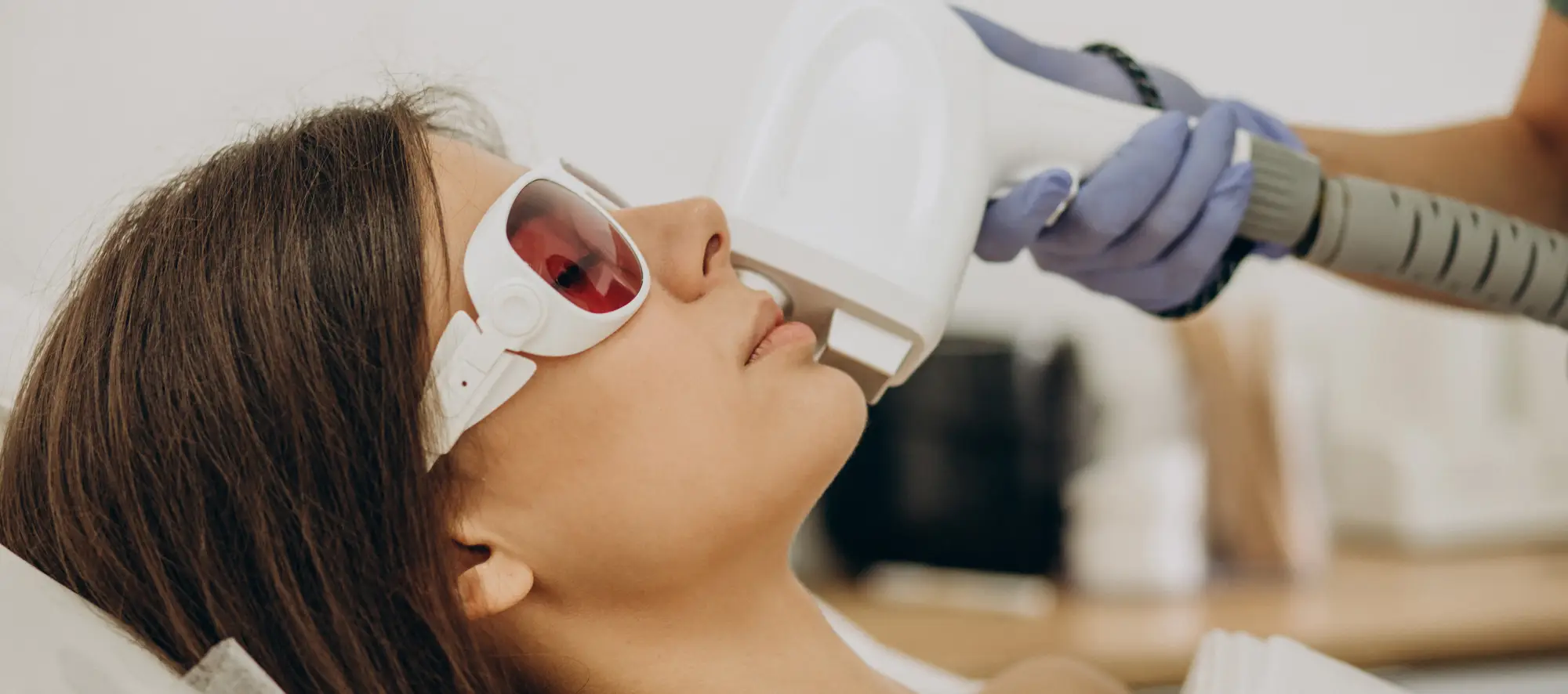
Pilot Heal boasts a team of highly skilled ophthalmologists with expertise in various retina-related surgeries, including retinal detachment. Our dedicated eye specialists are committed to preserving healthy vision for every patient. We operate our own clinics and collaborate with esteemed hospitals across India to ensure accessible treatment. Our state-of-the-art facilities are equipped to diagnose and manage a wide range of eye conditions. At Pilot Heal, retinal detachment is treated using various methods, including:
- Laser surgery and freezing
- Pneumatic retinopexy
- Scleral buckling
- Vitrectomy
With Pilot Heal, you can trust that you'll receive top-notch retinal detachment treatment at affordable prices. Contact us to schedule a consultation with one of India's finest ophthalmologists.
How is retinal detachment treated?
Diagnosis
Diagnosing retinal detachment involves a comprehensive eye examination to assess visual acuity, eye pressure, eye appearance, color perception, and the retina's ability to transmit signals to the brain. Additionally, the blood supply to the retina and its condition are evaluated. Imaging tests may also be recommended to visualize the retina and determine the extent of detachment. These imaging scans include:
Optical coherence tomography (OCT): A non-invasive and painless procedure that uses light waves to create 3D color-coded cross-sectional images of the retina.
Ocular ultrasound: Although it may cause slight discomfort, eye ultrasounds are typically performed with numbing eye drops. The procedure involves applying ultrasound gel to closed eyelids and scanning for digital imaging.
How to prevent retinal detachment?
- While there is no foolproof method to prevent retinal detachment, you can take certain precautions to reduce the risk:
- Use protective eyewear during sports and heavy lifting, particularly in activities with a higher likelihood of head or eye injuries.
- Wear specialized goggles and protective eyewear when handling power tools.
- Maintain proper blood sugar control if you have diabetes and manage other systemic conditions.
- Prioritize regular eye examinations and promptly consult an eye specialist when experiencing symptoms.

